Tallinn University
Natural and exact sciences
Molecular Biochemistry and Ecology
Maria Gnidenko
Capillary electrophoresis
Essay Supervisor: Kert Martma
Tallinn
2015
Table of contents
Acronyms and
symbols used
Introduction History and
development Physical basis and principle of separation
Elektrophoresis
Electroosmotic flow
Separation
process Electrodispersion
Various methods of separation
Capillary zone electrophoresis (CZE)
Micellar Electrokinetic Capillary Chromatography (MECC OR MEKC)
Capillary Gel Electrophoresis (CGE)
Capillary Isoelectric Focusing (
CIEF )
Isotachophoresis (ITP)
Electrokinetic Chromatography (EKC)
Micro Emulsion Electrokinetic Chromatography (MEEKC)
NonAqueous Capillary Electrophoresis (
NACE )
Capillary Electrochromatography (CEC)
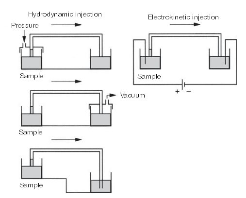
Equipment Sample injection
Electrokinetic injection (EI)
Hydrodynamic injection (HI)
Stacking
Capillary
Consitioning
Thermal
regulation High
voltage power source
Detection
Absorption in the UV and
visible region Notes to the quantitative
analysis 1
CE equipment
CE
application CE application in pharmaceutical analysis
CE application in biopharmaceutical analysis
CE application in biotechnology
Conclusions and
perspectives New directions
References
Acronyms and symbols used
API
Active pharmaceutical ingredients
CE
Capillary electrophoresis
CIEF
Capillary Isoelectric Focusing
CZE
Capillary zone electrophoresis
EI
Electrokinetic injection
EOF
Electroosmotic flow
GC
Gas Chromatography
HECE
High Efficiency Capillary Electrophoresis
HPLC High
Performance Liquid Chromatography
MEKC
Micellar Electrokinetic Capillary Chromatography
UV
Ultraviolet Introduction
Сapillary electrophoresis is a relatively new
method of analysis, which,
however , is rapidly
develops. It is
suitable for separation of polar and ionic samples and complements the
classical separation techniques
such as HPLC (High Performance Liquid Chromatography)
and GC (Gas Chromatography). All electrophoretic separation methods are
based on the
principle of
different rates of
migration of charged
particles and molecules in a
constant electric field .
2
CE (Capillary electrophoresis) combines different techniques of electrophoresis and
chromatography to
individual method that provides separation with high r
esolution i n a short
time. Additional commercial equipment for CE
offers the possibility of automatic sample
introduction, micropreparative fraction
collection , computer
control and data collection, as
well as the
registration of samples by Ultraviolet (UV)fluorescent, electrochemical or
radiochemical detectors (Беккёр, 2009).
History and development
History of the electrophoresis started in 1807, when
professor of the Moscow U
niversity, F
.
Reis
discovered phenomena such as electroosmosis and electrophoresis. However, the
practical use of this process in biology and
medicine b
egan much l ater and is a
ssociated w
ith
the name of the chemistry Nobel Prize winner
Arne Tizelius, who in the 30s
year of the last
century has
developed a method of electrophoresis in free fluid and constructed a
device for
electrophoretic separation a
nd a
nalysis of p
rotein m
ixtures b
y f
ree o
r
moving boundaries. The
main disadvantage of this method was the
heat liberation
during the electriс
current passing through the liquid b
ecause that prevented for
clear division o
f f
actions a
nd l ed to a
b
lurring of
the boundaries
between the individual
zones . In 1940, D. Philpot suggested to use columns
with a
density gradient of
buffer solutions, and in the 50s
years the method has been r
efined
and was developed the device for electrophoresis in density gradient.
However, the method was deficient, because after switching off the electric current,
formed during electrophoresis zones have "blurred". Subsequent achievements in electrophoresis
associated with the stabilization establishment of zones in the
solid support
medium . So, in
1950, as a solid carrier scientists began to use a
filter paper, in 1955 it was proposed to use
starch, and
already in 1957,
Cohn proposed to use as the solid support the
films of cellulose
acetate , which still remains one of the most
commonly used carriers at clinical
studies .
Almostly the
same time has been developed a method in which agarose is used as the basis.
In 1960 have been developed a method of capillary electrophoresis and only in 1989 was
created and put into
practice the
first analyzer, based on capillary electrophoresis method
(Шевченко et al., 2006).
3
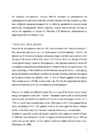
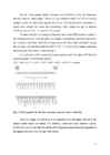
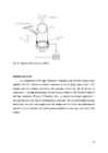
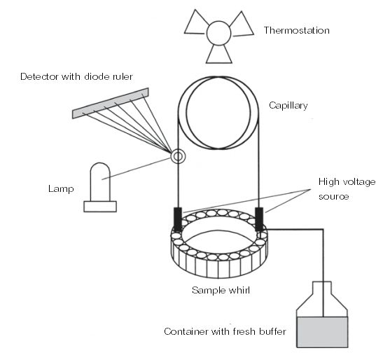
Physical basis and principle of separation
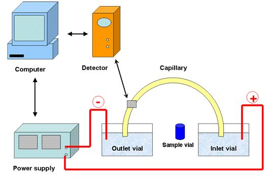
The principle of separation in CE is based on the different electrophoretic migration
of substances in an electric field. Principle of CE devices is v
ery
simple a
nd is s
hown i n F
ig.
1. Analysis sequence i s also very s
imple.
Both ends o
f t he capillary with e
lectrodes i mmersed
in the electrolyte container. The c
apillary i s f
illed with b
uffer, t o which the s
ample
solution i s
applied on one side. Then, sample
components are separated by an applied voltage, as they
move in a buffer at different speeds.
Fig. 1: Diagram of capillary electrophoresis system
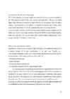
The principle of electrophoresis is strictly obeys Ohm's law. For the electrophoretic
separation must be keeped a constant voltage. The buffer is needed t o
provide an electrolytic
conductivity. In capillary convection and the free flow are minimized.
Therefore ,
highperformance capillary electrophoresis can be carried out in the same aqueous buffer.
Without polyacrylamide or agarose as a supporting matrix for electrophoresis, separation of
substances
takes place in f ree solution, i .e. in t he "open" c
apillary. S
o in t his
case w
e speak o
f
socalled free zone electrophoresis.
Necessary for this separation
technology devices must
4
create a field
strength of 200 to 400 V / cm to provide sufficient ion
mobility . Voltage,
depending on the capillary
length ,
lies between 5
a
nd 2
5 k
V.
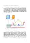
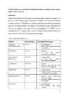
Typical operating p
arameters o
f
capillary electrophoresis are presented in Table. 1.
Table 1: Typical parameters of CE
Inner
diameter of the capillary
20100 μm
Capillary lenght
100150 cm
Voltage across the capillary
1030 kV
Current
rate 10100 μA
Field force
100500 W/cm
Generated heat
0,55 W
Sample
volume 150 nL
CE has one
advantage over
column chromatography
processes . It is not necessary to
wait
until the last
peak will cover the
distance to the
exit of the capillary, as after each
analysis, the capillary is washed with a special solution and
filled with new buffer.
Because of the use of quartz capillary filled with buffer, onlineUV detection at
wavelengths up to 200 nm is possible. The advantage of
direct passage of a beam of
light through the capillary is the
fact that in this case
there is no
band broadening caused by the
detector cell and inlet capillaries.
This detection does not introduce additional dead space. Its
drawback , however, is a small
internal diameter of capillaries (from 25 to 100 micrometers). Such a small optical
path length, according to the
Beer Lambert law,
results only a small absorption. Because of this
constructive specificity,
particular detection i n CE, c
ompared t o
other separation p
rocesses, is
insensitive method.
Elektrophoresis Electrophoretic separation is based on different rates of migration in an electric field, at that
the ion
velocity
v is the p
roduct o
f its electrophoretic mobility μ and an applied electric f ield
с
E:
v= μ
*E, where
electrophoretic mobility; E the a
pplied e
lectric
с
v internal velocity; μ
field.
5
The electric field is a
function of the applied voltage and t he length of the c
apillary w
ith the
dimension of V / cm. Electrophoretic mobility of the ion in the medium is a
c
onstant which
is calculated by the equation: μ
c =
q ,
6πη
rwhere q ion
charge ; η viscosity of the solution; r the radius of the ion
Small highly charged particles have high mobility; large particles having a low charge, in contrast , have a low mobility. Moreover , the electrophoretic mobility is
dependent on the pH of the solution. The
electrophoretic mobility is shown in tables as a physical constant defined on
complete dissociation of the electrolyte and extrapolation to infinite dilution. H
owever, in p
ractice it is
often substantially different, because the socalled effective mobility, in many
cases highly
dependent on the pH and
composition of the used buffer.
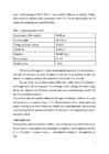
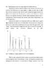
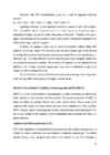
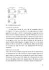
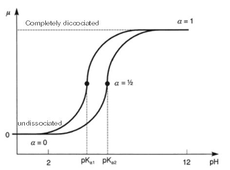
The distinction between absolute and effective mobility is explained in Fig. 2. There are
shown two hypothetical solution, which on c
omplete dissociation o
f t he electrolyte, i .e. w
hen
α (degree of dissociation)=1 have the same mobility μ.
Fig. 2: Mobility of two
weak acids as a function of pH
6
According to the absolute mobility, taken from the tables, both of
these substances seem
inseparable because they migrate e
qually. H
owever, b
oth these s
ubstances have d
ifferent p
Ka
values, i.e mobility is dependent on pH.
Electroosmotic flow (EOF) Migration of substances depends on the one
hand , on specific for compounds parameters as
charge and
size , and on the other hand, it is
affected by factors s
uch as pH and ionic strength
of the
working buffer, the field strength and temperature. Migration is complicated by
electroosmotic flow w
hich i s caused b
y c
harges o
n t he inner surface of t he capillary. EOF is a
feature of a capillary electrophoresis and is not
observed in conventional e
lectrophoresis i n a
flat layer.
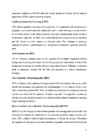
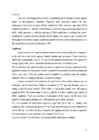
Electroosmotic flow is directed to the cathode stream of buffer. 7
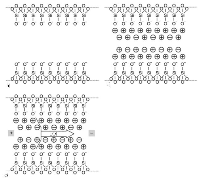
Fig. 3:
The appearance of osmotic flow: a) negatively charged surface of the quartz; b)
hydrated cations adsorbed on the surface; c) solution migrates to the cathode due to electric
field appearance
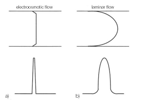
Fig. 4: The profile of and corresponding profile of the chromatographic zone
In aqueous solution, the majority of solid s
urfaces h
ave excess n
egative charge w
hich
appear due to ionization of the surface due to a
cid
base equilibrium a
nd / or a
dsorption of t he
ionized particles on the s
urface. In the q
uartz c
apillaries c
an o
ccur both, h
owever E
OF mainly
caused by dissociation of silanol groups (SiOH), which exist in the anionic form SiO
, as
shown in
Figure 3a.
To neutralize the charge counter ions are adsorbed on the surface and form an
electrical double layer, which
causes a potential difference, a socalled ζpotential (Figure
3b). If now apply a voltage, the cations of the diffuse double l ayer m
igrate i n the d
irection o
f
the cathode.
Since cations are solvated, the solution
around them moves
along with them in
the direction of the cathode (Figure 3c).
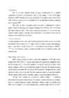
This electroosmotic flow does not obey to the l aw o
f HagenPoiseuille about t he f
low
in a capillary under
pressure . It differs from it by a very flat flow profile. Since the driving
force along the capillary (i.e on i ts i nner w
alls), i s h
omogeneous, the p
ressure drop a
cross t he
8
capillary is absent. This
effect explain a very slight broadening of the bands observed in the
CE (Fig. 4).
Separation process Band separation in zone electrophoresis is based on a combination of electrophoretic mobility and electroosmotic flow of ions. In electroosmosis effect voltage
value , ionic strength, viscosity of a buffer, additives
in
eluent and different coatings of the capillary walls. Electrophoretic mobility of
positive ,
neutral and
negative sample molecules is different, but all of the particles under t he effect o
f
electroosmotic flow migrate towards the cathode.
The rate of migration of the particle is the sum of its own electrophoretic mobility and electroosmotic flow rate. The advantage of electroosmotic flow is the fact that
almost all the particles are
moving independently of the charge in the same d
irection. I
n normal conditions, i.e. c
apillary
surface is negatively charged, the flow moves from the anode to the cathode. The anions are
also moving to the cathode, as EOF may is measurable
greater than the electrophoretic
mobility. That
means , that cations, anions and neutral molecules can be separated
electrophoretically in a
single analysis since they migrate in the same direction.
● Cations moved most quickly, as the EOF and electrophoretic
movement directed in
the same direction to the cathode
● The neutral molecules are transported at a
speed of EOF, but not separated.
● Anions migrate more slowly as they are attracted to the anode, but move under the
influence of EOF
toward the cathode.
Migration time is
determined by a characteristic constant of ion m
obility a
nd e
lectroosmotic
flow coefficients:
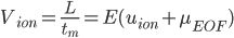
,
where V velocity migration; L
length of c
apillary (
the detector); t t ime of m
igration; E
m
electric field strength; u ion mobility; μ the coefficient of EOF.
ion
EOF
In the analysis of small ions (e.g., N
aCl, KCl), their m
obility i s u
sually s
o l arge t hat i s
comparable to the EOF. An additional surface modification of capillary EOF c
an be reduced
9
to such an extent that the movement of anions and cations in setting direction will stop.
Conventional
coating of capillaries surface with
linear polymers without transverse
crosslinks eliminates electroosmosis, which is typical for uncoated capillaries.
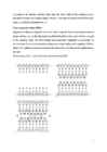
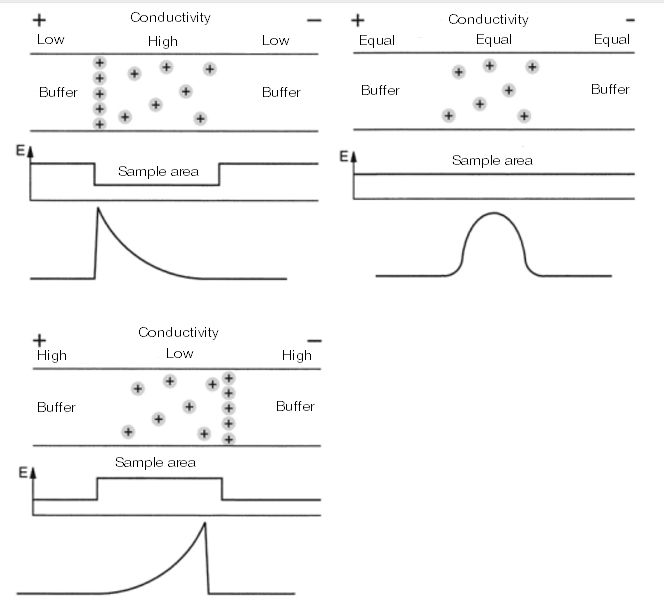
Electrodispersion The peculiarity of capillary electrophoresis is the electrodispersion (Figure 5).
Because of this can be
seen as symmetric
Gaussian and triangular asymmetric peak shapes.
While the distorted shapes of the peaks with "
front " and "
tails " in the other c
hromatographic
process can be eliminated by adoption of appropriate measures, such peaks shapes in CE
should be
considered as normal. T
hey a
re caused b
y t he
strong differences in t he conductivity
of the sample and buffer.
If the sample has a
higher mobility than the separation buffer, the front part of the zone
becomes diffusive, and the
tail of the band becomes sharper ("front" peak). On the contrary,
at a lower mobility of the sample, than a buffer received acute frontal z
one and diffusive e
nd
zone ("tail" of the peak). Symmetrical peaks are observed only if both the conduction are
identical.
10
Fig. 5: Electrodispersion caused by varying conductivity of buffer and sample
Unsymmetrical peaks are caused by different conductivity and, at the same time, by
an electric field. If the sample has a higher mobility, i.e. higher conductivity than t he buffer,
then on the both sides (on the front, and on the end of sample zone)
appears excessive field
gradient in the transition zone. The
stress gradient in the end zone of the sample is directed
the same as the migration of t he sample, s
o t hat t he sample c
omponents in t ransition z
one are
accelerated
back towards the transition z
one. B
ecause of t his, a
ppears a
s
harp boundary a
t the
end of the peak sample.
Conversely, the particles in the
forward zone of the sample diffuse into the region of
high field gradient, such that they are accelerated more in the same direction. Consequently,
the front zone is drawn farther so that there is a "fronting" o
f peak. The opposite effect
peak
"tailing" occurs if the mobility of sample area i s less than t he mobility o
f r
unning b
uffer. O
n
neutral
particles
these
conductivity
differences
do
not
affect .
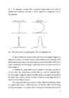
Fig. 6: "Fronting" and "tailing" of peaks as a
result of electrodispersion
While this peaks asymmetry are always
present , it is in most cases s
maller c
ompared
with other dispersion effects, such as
diffusion . On Fig. 6 are depicted a
nions t hat a
re highly
distinguished by their mobility. It is
known that the "fronting" is observed in
fast eluting
11
anions with high mobility, Gaussian peaks at an
average mobility of anions and "tailing" is
observed in lowmobility anions which eluted last.
If it does not interfere to peak resolution, this asymmetry can b
e n
eglected. H
owever,
it can also b
e e
liminated b
y b
ringing t he conductivity of w
orking b
uffer t o the c
onductivity o
f
the sample.
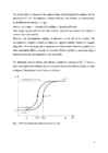
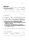
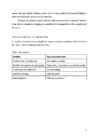
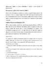
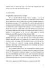
Various methods of separation
In capillary electrophoresis are manifested various separation mechanisms that are listed in
the Table. 2 and schematically depicted in Fig. 7.
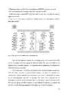
Table 2: CE methods
Methods Separation mechanism Capillary zone electrophoresis
Zone solution mobility
Micellar electrokinetic chromatography Hydrophobic / ionic interaction with the micelles
Capillary gel electrophoresis
Size and loading
Isoelectric focusing
Isoelectric point
Isotachophoresis
“Moving connections”
12
Fig. 7: A schematic representation of zone electrophoresis, isoelectric focusing and
isotachophoresis
Capillary zone electrophoresis (CZE) Because of their
ease of use and versatile applications CZE is the most commonly
used method which is primarily u
sed f
or t he separation o
f small w
ater
soluble m
olecules. I
t i s
used in the analysis of amino acids, peptides, and ions of various enantiomers (optically
active compounds) and many other ionic compounds. CZE it is t he simplest f
orm o
f HECE
(High Efficiency Capillary Electrophoresis) as capillary f
illed only w
ith buffer. Separation o
f
materials into discrete zones is due to migration at different speeds.
The electroosmotic flow (EOF)
makes possible the separation of both cations and
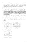
anions. Neutral molecules do not migrate electrophoretically and move with EOF.
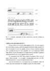
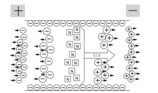
Fundamental processes of capillary electrophoresis shown in Fig. 8.
Since the electroosmotic flow can be greater than the electrophoretic mobility, the
cations, anions and neutral particles move through the capillary with different speeds.
13
• Cations moved most quickly, as the electrophoresis and EOF have the same direction.
• The neutral particles do not separate, they move only under the EOF.
• Small anions migrate against EOF toward the anode, because t heir electrophoretic mobility
higher than the EOF.
• Large anions slowly migrate towards the cathode because their electrophoretic mobility
lower than the EOF.
Fig. 8:
The principle of capillary zone electrophoresis
Since the electrophoretic mobility of ions depends on the pH, the selectivity in CZE
can be very simply varied by changing the pH of the buffer. The type of the buffer is very
important for a successful separation. The separation can be optimized by the
addition of a
surfactant or chiral components.
CZE is o
ften u
sed in t he biology, p
articularly f
or the a
nalysis of p
eptides and proteins.
CZE also finds application in pharmaceutical analysis, the study of metabolites and
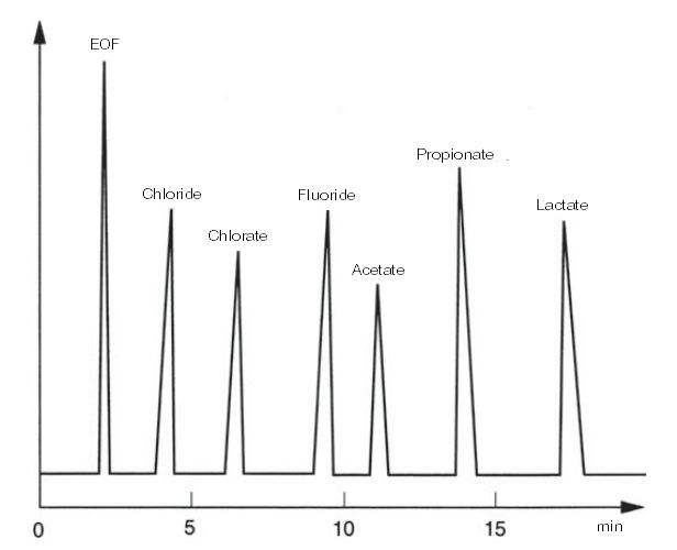
environmental analysis. Inorganic ions and
organic acids, which are traditionally determined
by ion chromatography, may also be analyzed CZE. Since ions, in general, are not
chromophores and
even absorb ultraviolet light, it is necessary to operate with
indirect UV
detection. For this use as buffer solutions are used solutions of chromate or imidazole and
detected at a buffer maximum absorption wavelength (e.g., 254 nm for chromate).
14
Ions of eluting sample displace chromate ions and thereby
reduce the absorption,
whereby "negative" peaks appear. Due to the high mobility of small ions EOF is not s
trong
enough to move the ions in the opposite direction of their electrophoretic movement, i.e.
anions move toward the anode and respectively, small anions can not be analyzed
simultaneously with other components of the sample.
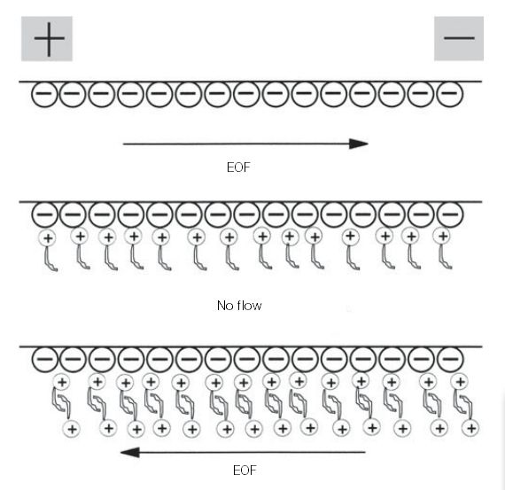
To reduce the EOF or to
change its direction, the s
ocalled E
OF m
odifier i s a
dded to
the working electrolyte. That role
plays , for example, a hydrophobic quaternary ammonium
salt or cationic surfactants, which due to adsorption on the silica s
urface can modify e
xisting
therein charge ratio and thus modify the EOF [7.141]. The
action of the cationic surfactants,
adsorbed on the silica surface is shown in Fig. 9.
In 5 millimolar (mM) solution of chromate migration used for indirect U
V d
etection
is generally presetn the
following sequence:
S
О 2–


Kõik kommentaarid