Utilizing data on the candidates' use of various social media sites, survey data from the 2011 Finnish election study, and survey ... Cited by 20 Related articles All 2 versions Cite Save 11. Kas leiate Krista Lepiku poolt 2014. aastal kirjutatud tekste? Kui jah, palju ja mida leiate? Jah leidsin 3 teksti. Kasutasin scholari täielikku otsingut. Return articles authored by: Krista Lepik Vajutasin otsi, siis edasi valisin since 2014 Antinuclear antibodies in atopic dermatitis: a cross‐sectional study on 346 children Nimi:ANNERIIN TRUU Õpperühm:IK 12 …, K Metsküla, T Annus, U Putnik, K Lepik… - International journal …, 2014 - Wiley Online Library Background Atopic dermatitis (AD) is a chronic inflammatory skin disease that can be classified into an extrinsic or intrinsic type. A high percentage of patients, especially adults with the extrinsic type of AD, have been reported to show antibodies to antinuclear ...
RA haigestumus · Sooline erinevus · Östrogeen TNF RA patogenees · Autoimmuunhaigus, võtmeprobleem T- lümfotsüütide tolerantsuse düsregulatsioon · Autoantikehade teke aastaid enne artriidi avaldumist · Anti-CCP (antibodies to cyclic citrullinated peptides) filagriini, keratiini, fibrinogeeni, vimentiini vastu · RF (rheumatoid factor) IgG vastu · 75% IgM · Sünoviaalkest hüpertrofeerub, tekib pannus granulomatoosne kude, mis on tekitatud fibroblastide proliferatsioonist ja milles on arvukalt põletikuliste rakkude infiltraatidega ümbritsetud väikesi veresooni. · Luu kahjustus kujuneb hiljem
Microscopy may be carried out with simple instruments, such as the compound light microscope, or with instruments as complex as an electron microscope. Samples obtained from patients may be viewed directly under the light microscope, and can often rapidly lead to identification. Microscopy is often also used in conjunction with biochemical staining techniques, and can be made exquisitely specific when used in combination with antibody based techniques. For example, the use of antibodies made artificially fluorescent (fluorescently labeled antibodies) can be directed to bind to and identify a specific antigens present on a pathogen. A fluorescence microscope is then used to detect fluorescently labeled antibodies bound to internalized antigens within clinical samples or cultured cells. This technique is especially useful in the diagnosis of viral diseases, where the light microscope is incapable of identifying a virus directly. Biochemical tests
Dopamine, epinephrine, norepinephrine, Pharmaceutical, urine ICL catechol CE application in biopharmaceutical analysis CE has demonstated to be a complementary alternative to chromatographic techniques in biopharmaceutical analysis in biopharmaceutical analysis. The complete characterization of biopharmaceutical drugs such as erytthropoietin and various therapeutic monoclonal antibodies glycosylation compositions, igG purity, and impurities for quality control purposes are of utmost importance. Further, therapeutic biomolecules have a higly complex composition and structure. Moreover, the products may vary in structure due to the complexity of cell culture and purification processes. Various modes of CE offer several
heparin) was obtained by a trained phlebotomist 10 min before the TSST and again 50 min after the TSST. The whole blood was centrifuged (250g for 10 mins) and the resulting plasma layer was removed, aliquoted and stored at 70 oC for cytokine analysis. All samples were numerically coded (known to the principal investigator) and laboratory measurements (see below) undertaken blind. 2.3.1 Detection of Plasma IL-6 Plasma IL-6 was detected using commercially available paired antibodies enabling cytokine detection in an ELISA (Enzyme Linked ImmunoAssay) format (R & D systems Ltd, Abingdon, UK). The sensitivity for the IL-6 ELISA was 9 1000 pg/ml. There was no reported cross-reactivity with other cytokines (R & D systems Ltd, Abingdon, UK). Emotion regulation in relation.. 1 2.3.2. Detection of Salivary Cortisol Salivary cortisol was dectected using a commercially available ELISA (Salimetrics, LCC, USA)
varem nende antigeenide suhtes sensibiliseerunud isikutel, kellel on vastavad IgE- klassi antikehad. 1. Süsteemne: anafülaktiline sokk- raske, generaliseerunud (mitut elundkonda haarav) ülitundlikkusreaktsioon. Kujuneb mõne minutiga, vajab kohest esmaabi (adrenaliini süst- kitsendab veresooni, avab hingamisteed, kõrgendab vererõhku, alandab sügelust). 2. Lokaalne: allergiline nohu, astma, toiduallergia A type I hypersensitive reaction is mediated by IgE antibodies, whose Fc region binds to receptors on mast cells or blood basophils. Crosslinkage of the fixed IgE by allergen leads to mast cell or basophil degranulation with release of pharmacologically active mediators. The principal effects of these mediators are smooth-muscle contraction and vasodilation. Tüüp II: tekivad enamasti autoantikehade mõjust kudedele, harvem on need tingitud mikroobidevastastest antikehadest. Koekahjustus tekib
sticky ends / described; ref. complementary sticky ends; ligase seals (sugar-phosphate) backbone / AW; max 4 (ii) credit any two from the following: 1 antibiotic resistance (gene) introduced and survivors have plasmid; 2 fluorescent marker (gene) introduced and glowing bacteria have plasmid; 3 identify bacteria producing insulin using antibodies; 2 [6] 8. referring to pig insulin: ethical / religious, reasons; incompatibility / lack of tolerance / immune response; ora not exactly the same as / less effective than, human insulin; ora referring to human insulin from bacteria: engineered insulin is cheaper; ora
and the remaining exons connected to re-form a single continuous molecule. Although most RNA splicing occurs after the complete synthesis and end-capping of the pre-mRNA, transcripts with many exons can be spliced co-transcriptionally.[5] The splicing reaction is catalyzed by a large protein complex called the spliceosome assembled from proteins and small nuclear RNA molecules that recognizesplice sites in the pre-mRNA sequence. Many pre-mRNAs, including those encoding antibodies, can be spliced in multiple ways to produce different mature mRNAs that encode different protein sequences. This process is known as alternative splicing, and allows production of a large variety of proteins from a limited amount of DNA. 26. Geneetiline kood http://et.wikipedia.org/wiki/Geneetiline_kood Geneetiline kood- on vastavus, kus mRNA kolm järjestikust nukleotiidi (st. koodon) määravad ära ühe aminohappe paigutuse valgu molekulis. 27
and the remaining exons connected to re-form a single continuous molecule. Although most RNA splicing occurs after the complete synthesis and end-capping of the pre-mRNA, transcripts with many exons can be spliced co-transcriptionally.[5] The splicing reaction is catalyzed by a large protein complex called the spliceosome assembled from proteins and small nuclear RNA molecules that recognizesplice sites in the pre-mRNA sequence. Many pre-mRNAs, including those encoding antibodies, can be spliced in multiple ways to produce different mature mRNAs that encode different protein sequences. This process is known as alternative splicing, and allows production of a large variety of proteins from a limited amount of DNA. 26. Geneetiline kood http://et.wikipedia.org/wiki/Geneetiline_kood Geneetiline kood- on vastavus, kus mRNA kolm järjestikust nukleotiidi (st. koodon) määravad ära ühe aminohappe paigutuse valgu molekulis. 27. Translatsioon, tRNAde ja ribosoomide ehitus
·Lüsosüüm-mukolüütiliselt toimivat leeliseline ensüüm. ·Teda leidub: süljes soole ja nina-neelu ruumi limas, silmalaugude sidekesta sekreedis ja pisarates granulotsüütide graanulites, kopsude makrofaagides, OPSONISATSIOON ·Opsiniinid on ained, millede seondumine keha võõraste rakkude ja mikroorganismidega muudab nad kergemini fagotsüteeritavateks ·Opsiniinid: antikehad, komplementfaktorid jt. Näiteks, antibodies: IgGand IgA components of the complement system: C3b, C4b, and iC3b Surfactant Mannose-binding lectin(initiatestheformationofC3b) The mos timportant are IgG and C3b. ·Nii fagotsüüdi kui ka võõrkehade pind on enamasti negatiivselt laetud.Seega on nende lähenemine raskendatud elektrostaatilise tõukejõu tõttu. Opsonisatsioonil seonduvad võõrkeha pinnale antikehad ja (või) komplemendi valgud. Fagotsüüdi pinnal on retseptorid, mis hõlpsasti seonduvad opsiinide molekulidega
otsest immunofluorestsentsmeetodit, milles uuritavaks materjaliks on koelõik. Määratakse antigeeni olemasolu ja lokalisatsiooni koelõigus, nt glomerulonefriit – neerubiopsia külmutuslõigul määratakse neerupäsmakeses immuunkomplekside olemasolu, lokalisatsioon ja koostis (Ak isotüüp); kaudset immunoflorestsentsmeetodit: määratakse järgnevaid autoantikehi – ANA e tuumavastased antikehad; SMA e anti-smooth muscle cell antibodies; nii ANA kui ka SMA võib leida ka tervetel inimestel, seetõttu nende leid ei viita veel väga kindlale haigusele. ELISA: antigeenidena kasutatakse nt ssDNA-d - SEL puhul on spetsiifilisus >90%, reumatoidartriidi puhul 40%; kardiolipiini – SEL puhul spets. 40% Skriining poolkvantitatiivse latekstestiga - määratakse reumatoidfaktorit (fS-RF) – see on oma olemuselt heterogeenne autoantikehade grupp, mis tekib IgG-molekuli Fc-fragmendi vastu. Need
higher shear force and/or less favorable ten- 2001; Lukoyanova et al. 2002). This region derness scores (Huff-Lonergan et al. 1995, of nebulin also has been suggested to inhibit 1996a, b). The T2 polypeptide can also be the sliding velocities of actin filaments over subsequently degraded or altered during myosin. If the latter role is confirmed, then it normal postmortem aging. Studies that have is also possible that nebulin’s postmortem used antibodies against titin have been shown degradation may alter actin-myosin interac- to cease to recognize T2 after prolonged tions in such a way that the alignment and periods of postmortem storage or μ-calpain interactions of thick and thin filaments in digestion (Ho et al. 1994; Huff-Lonergan postmortem muscle is disrupted. This, too, et al. 1996a) could lead to an increase in postmortem ten-
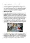
This must not be confused with the normal sprinkling of isolated white hairs which appear in a cat's fur during its lifetime. "Leukotrichia" is a generic term. Acommon term is "piebaldism" because it causes white patches in the skin and fur. It is an "aquired depigmentation" that occurs during the cat's lifetime, is usually progressive and may be triggered by illness or environmental factors. Ultimately, a cat with leukoderma may become almost entirely white. Antibodies are formed against the pigment-producing melanocytes. The melanocytes are destroyed leading to the white areas. A type of leukoderma has been identified in some Persian cats and these are used as laboratory subjects in the study of depigmentation conditions. Periocular leukotrichia, causes the fur around the cat's eyes to become pale - as though the cat is wearing spectacles. Vitiligo in a big cat - the "cobweb panthers" that turned progressively white.