0140108) and the Estonian Science Foundation (Grant No. 9185) (www.etis.ee). The funders had no role in study design, data collection and analysis, decision to publish, or preparation of the manuscript. Competing Interests: The authors have declared that no competing interests exist. * E-mail: [email protected] Introduction (based on the peak heights of different nucleotides in the same position on the electropherogram) have been focused on analysing Ribosomal RNA genes (rDNA) have been widely used in alleles that differ by nucleotide substitutions [5,6]. Whereas, if the taxonomy, biogeographic and phylogenetic analyses. A eukaryotic gene variants differ due to insertion or deletion events, and
accuracy of the estimation of differences at high levels of stimulus is much less important for survival. http://www.bact.wisc.edu/Microtextbook/modules.php? op=modload&name=Sections&file=index&req=viewarticle&artid=10&page=1 (LOE LÄBI!!!) 10. Miks geneetiline kood on tripletne? The genetic code is a triplet code, with three nucleotides coding for one amino acid. In other words, three nucleotides in mRNA (a codon) specify one amino acid in a protein. (DNA RNA valgud) 11. Kirjeldage, mida peetakse molekulaarbioloogia tsentraalseks dogmaks ja kuidas toimuvad replikatsioon, transkriptsioon ja translatsioon. Geneetilise informatsiooni ülekande suunda DNA RNA valk nimetatakse oma keskse tähtsuse tõttu molekulaarbioloogia põhidogmaks. 1. Replikatsioon - päriliku materjali (mis võib olla nii DNA kui RNA) kahekordistumine. Elusorganismide
The sigma factor greatly reduces the affinity of RNAP for nonspecific DNA while increasing specificity for certain promoter regions, depending on the sigma factor. That way, transcription is initiated at the right region. The complete holoenzyme therefore has 6 subunits: 2' (~480 kDa). The structure of RNAP exhibits a groove with a length of 55 Å (5.5 nm) and a diameter of 25 Å (2.5 nm). This groove fits well the 20 Å (2 nm) double strand of DNA. The 55 Å (5.5 nm) length can accept 16 nucleotides. When not in use RNA polymerase binds to low affinity sites to allow rapid exchange for an active promoter site when one opens. RNA polymerase holoenzyme, therefore, does not freely float around in the cell when not in use. 12. Joonista tüüpilise PolII promootori struktuur ja kirjelda faktoreid, mis sinna transkriptsiooni initsiatsiooni käigus seovad. Mediaatorkompleks ja selle tähtsus. 5'......TATAAA...3'
referring to human insulin from bacteria: engineered insulin is cheaper; ora greater supply of engineered insulin; ora 1 [1] 9. allow max 5 for following: transcription; DNA unzips / H bonds break; exposing required, gene / sequence of bases; RNA nucleotides align with DNA; U with A, A with T, C with G, and G with C; RNA polymerase; mRNA formed (using DNA strand as template); leaves nucleus through pore; allow max 5 for following: translation; mRNA attaches to ribosome; tRNA brings amino acid (to, ribosome / mRNA); each tRNA attached to specific amino acid; tRNA binds to mRNA using complementary, base triplet / anticodon; peptide bond formed between amino acids;
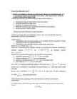
to the scoring system in use (BLAST 2.0 defaults). Gap: A space introduced into an alignment to compensate for insertions and deletions in one sequence relative to another. To prevent the accumulation of too many gaps in an alignment, introduction of a gap causes the deduction of a fixed amount (the gap score) from the alignment score. Extension of the gap to encompass additional nucleotides or amino acid is also penalized in the scoring of an alignment. d. Varieerida parameetreid ükshaaval, selgitada nende mõju tulemustele: Mõju avaldas parameetrite muutmine vaid skoorile, expect ei muutunud. i. Leida skoor ja E järjestuse gi|21325986|gb|AAM47554.1| jaoks järgmistel parameetrite kombinatsioonidel: >gi|21325986|gb|AAM47554.1|AF428108_1 alpha-enolase [Crocodylus palustris]
The diphosphate 5' prime end then attacks the gamma phosphorus atom of a GTP molecule in order to add the guanine residue in a 5'5' triphosphate link. The enzyme (guanine- N7-)-methyltransferase ("cap MTase") transfers a methyl group from S-adenosyl methionine to the guanine ring.[3] This type of cap, with just the (m7G) in position is called a cap 0 structure. The ribose of the adjacent nucleotidemay also be methylated to give a cap 1. Methylation of nucleotides downstream of the RNA molecule produce cap 2, cap 3 structures and so on. In these cases the methyl groups are added to the 2' OH groups of the ribose sugar. The cap protects the 5' end of the primary RNA transcript from attack by ribonucleases that have specificity to the 3'5'phosphodiester bonds.[4] [edit]3' Processing Main article: Polyadenylation [edit]Cleavage and Polyadenylation
The diphosphate 5' prime end then attacks the gamma phosphorus atom of a GTP molecule in order to add the guanine residue in a 5'5' triphosphate link. The enzyme (guanine- N7-)-methyltransferase ("cap MTase") transfers a methyl group from S-adenosyl methionine to the guanine ring.[3] This type of cap, with just the (m7G) in position is called a cap 0 structure. The ribose of the adjacent nucleotidemay also be methylated to give a cap 1. Methylation of nucleotides downstream of the RNA molecule produce cap 2, cap 3 structures and so on. In these cases the methyl groups are added to the 2' OH groups of the ribose sugar. The cap protects the 5' end of the primary RNA transcript from attack by ribonucleases that have specificity to the 3'5'phosphodiester bonds.[4] [edit]3' Processing Main article: Polyadenylation [edit]Cleavage and Polyadenylation
age, nutritional level of the animal, and connective tissue. muscle type. It is important to note that the There are numerous non-protein nitroge- lipid content varies inversely with the water nous compounds in skeletal muscle. They content (Callow 1948). Some lipid is stored include substances such as creatine and cre- inside the muscle cell; however, within a atine phosphate, nucleotides (ATP, ADP), muscle, the bulk of the lipid is found between free amino acids, peptides (anserine, carno- muscle bundles (groupings of muscle cells). sine), and other non-protein substances. Average lipid content of skeletal muscle is about 3% of the muscle weight, but the range Muscle Structure can be as much as 1–13% (U.S. Department of Agriculture 2008)